Case 24 - A Narrow Escape
Author: Dr Angus Perks Reviewer: Dr Nish Cherian
A 77 year old male presents after a fall and long lie. He has no PMH and has not seen a doctor in 10 years. He suddenly is noted to be less responsive and becomes pale, unresponsive and drops his RR to 8. You rush him to Resus where he regains consciousness and his colour improves.
You elect to perform a focused echo to assess why he may have transiently appeared peri-arrest: -
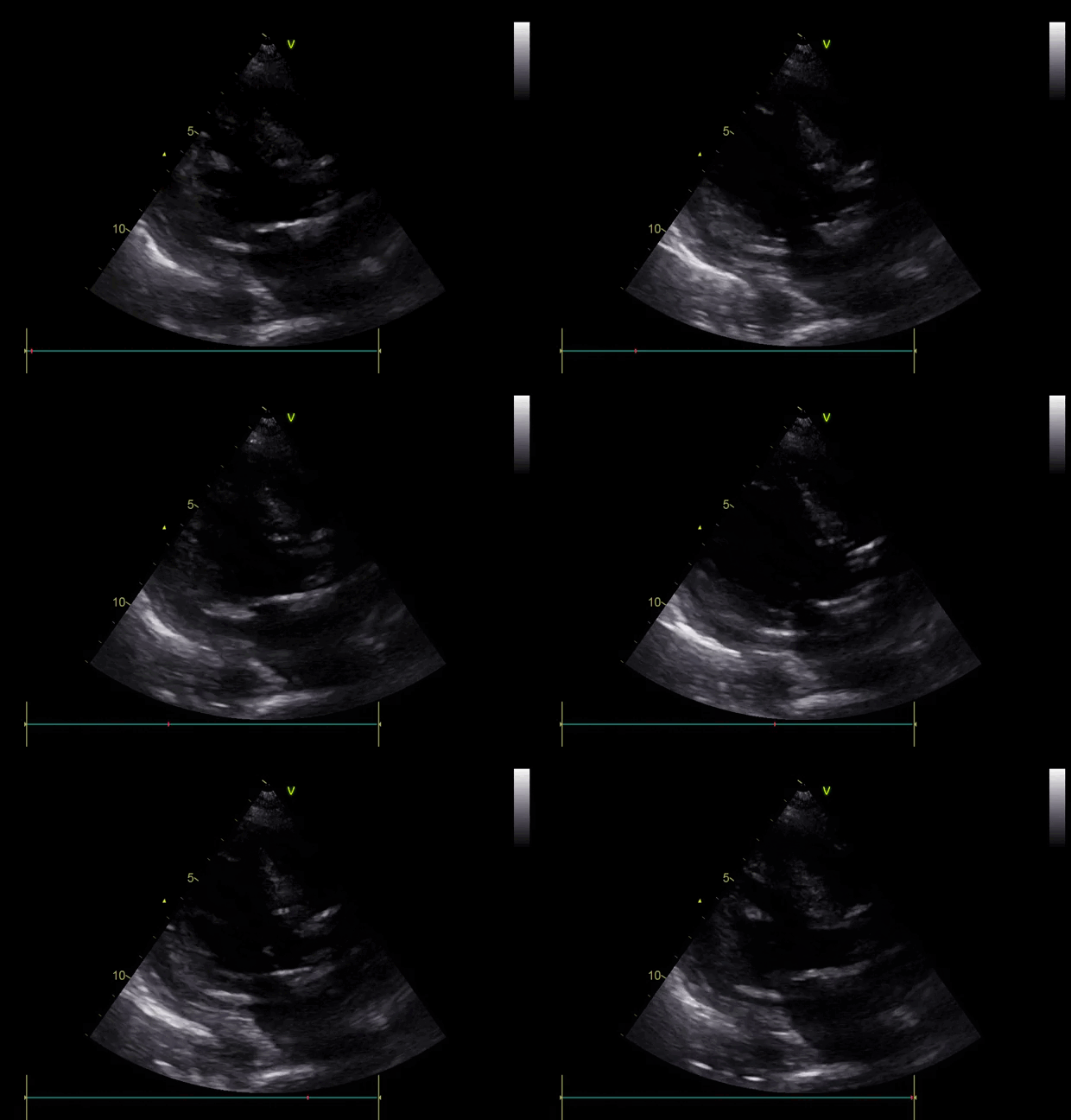
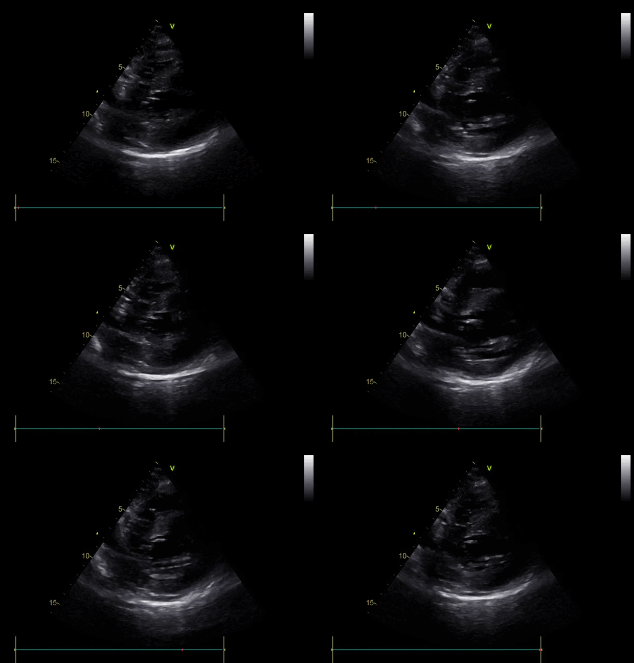
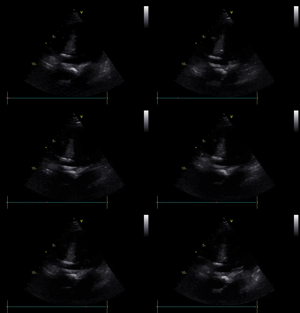
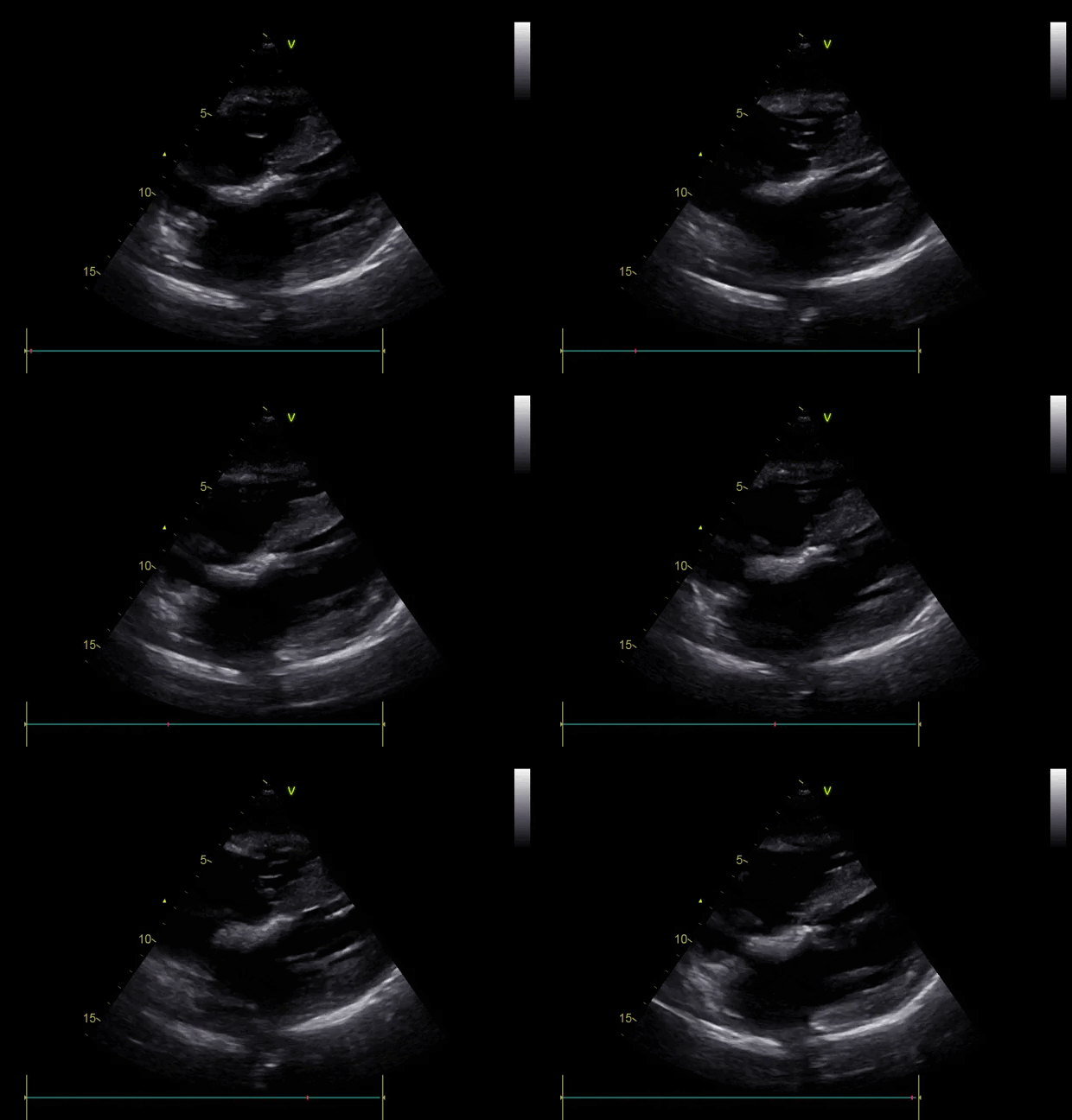
Clip Collection (Press right/Left to change the clips)
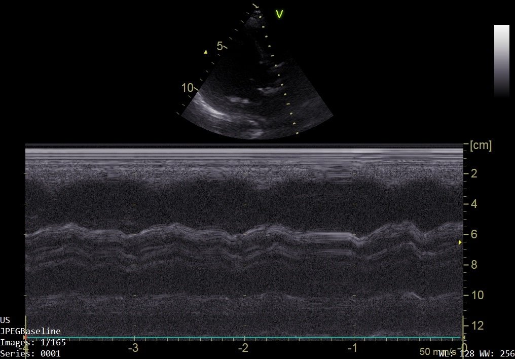
Focused TTE: PLAX, PLAX M-mode through AV, PSAX, A5Ch (zoomed) and SC
*TTE- Transthoracic Echocardiography, PLAX- Parasternal Long Axis, AV- Aortic Valve, PSAX- Parasternal Short Axis, A5Ch- Apical 5 Chamber, SC- Subcostal
-
This was a difficult study due to very low body habitus and patient positioning.
However, the following pertinent findings can be appreciated:
• There is notable LV Hypertrophy
• The AV appears heavily calcified and is hardly moving
-
This is highly suspicious for critical Aortic Stenosis, given the combination of echo findings and clinical history.
(N.B. a diagnosis of Aortic Stenosis cannot be made without a departmental comprehensive echo study as it requires detailed measurements. A focused study can raise suspicion though – these patients should then have a departmental echo to quantify findings.)
-
‘M-mode’ is a ‘time-motion-display’ of ultrasound along a chosen line. Basically, it shows ultrasonic reflections along the chosen line across time. It can sample at very high time resolution and frequency, allowing for assessment of rapid motion (e.g. cardiac valve leaflets).
Imagine it as a recording of the motion of all objects in that one ultrasound beam across time.
See below the comparison of a normal AV and our patient’s.
Look here at the reference image of M-mode through the AV (left). Contrast this through the image of our patient (right).
M-mode assessment of the AV in this patient demonstrates minimal leaflet opening.
CASE RESOLUTION
This patient was admitted under the care of Cardiology and went on to have a departmental echo confirming critical aortic stenosis.
Take Home points
With appropriate training, TTE can be used to assess for significant valvular pathology
Visual appearance of the valve and opening/closing is of equal importance to advanced measurements/gradients and will clue you in to a diagnosis or highlight need for a comprehensive/Level 2 scan.
Focused TTE can NOT make a diagnosis of valve stenosis, only raise suspicion and prompt referral for a comprehensive echocardiogram
FOCUSED ECHO TRaining COVERING VALVULAR ASSESSMENT
Current UK focused echo training that includes valvular assessment is either through: -
British Society of Echocardiography’s Level 1 accreditation
Intensive Care Society’s FUSIC HD accreditation


