Sound Bite 10- Pneumonia (Paeds)
Author: Dr Nick Mani
3yr old child presents to the local Emergency Department with cough, wheeze, fever. On examination, sats 90-91% despite nebs for viral wheeze/URTI, ?wheeze/crackles worse on the left side (lower>upper) on reassessment by a senior.
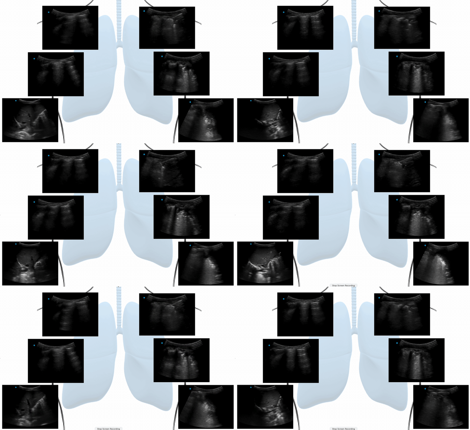
You as the senior elect to perform lung POCUS as shown below: -
Clips- Lung POCUS views (anterior). The right side A-lines present. The left side shows multiple B-lines (worse lower zone), some of which are coalescing (‘white-lung’), consolidation with air-bronchograms. The left lower base was tricky to scan, but there was no pleural effusion (the dot/left side of the screen is the head end of the patient)
Lung POCUS findings in the context of the presentation and standard clinical assessment is more likely to be due to left sided pneumonia. CXR was equivocal.
Case Resolution: -
The child was started on IV antibiotics, oxygen and admitted to the ward for ongoing assessment and management.
Take Home Message: -
Lung POCUS could be useful the diagnosis in children with respiratory symptoms when the diagnosis is still unclear. It could help to differentiate between viral and bacterial respiratory tract infection, and sequela such as effusion.

