
Case 2 - Don't flap!
Author: Nish Cherian Reviewer: Nick Mani
A 64-year old non-English speaking woman presents to ED at 2am with a sudden onset of chest and abdominal pain. She is agitated and writhing around on the trolley. She is hypertensive 180/70 and tachycardic 115bpm, but other vitals are normal.
You are concerned about the possibility of an acute aortic syndrome and there is at least a 2h wait for CT. POCUS shows the following:

Clips Collection-
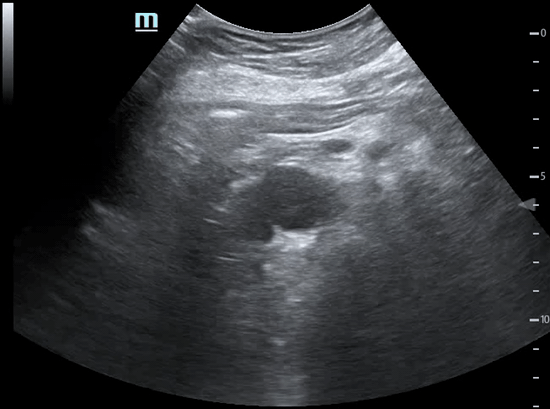
Clip 1a- Short Axis view of Abdominal Aorta
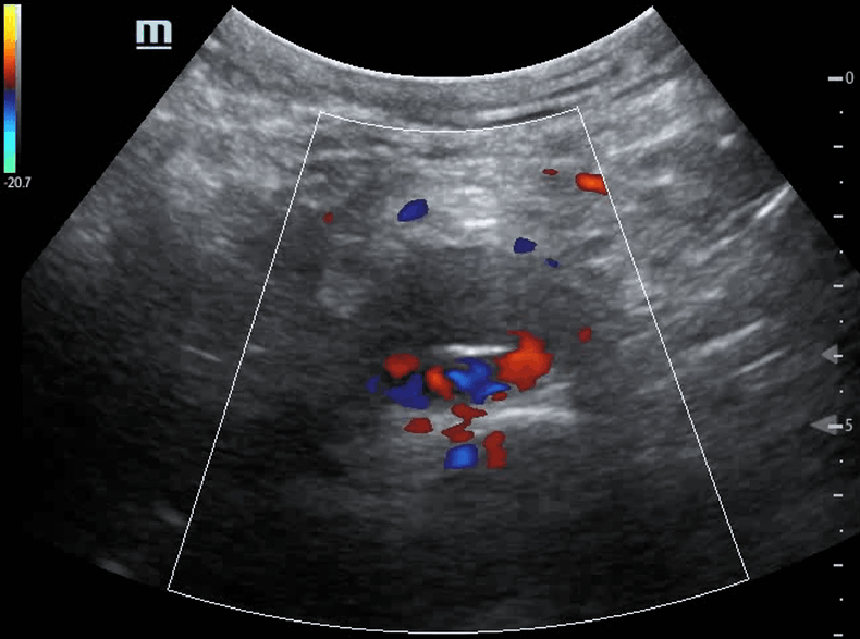
1b- Short Axis view of Abdominal Aorta, Colour Doppler on
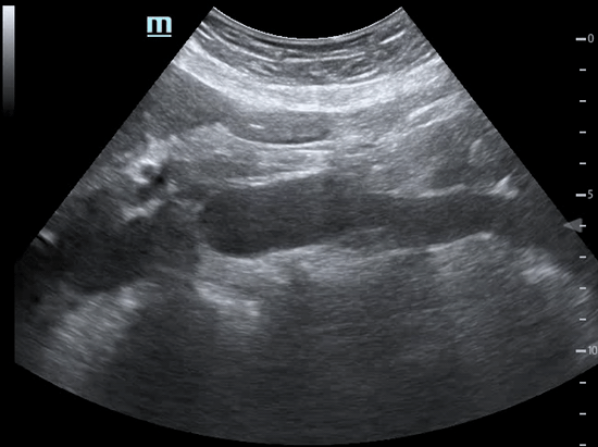
Clip 2a- Long Axis view of Abdominal Aorta
2b- Long Axis view of Abdominal Aorta, Subxhipoid
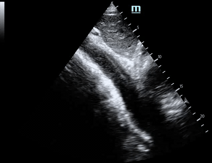
Clip 3a- Aortic Arch
3b- Aortic Arch, Colour Doppler on
-
An intraluminal intimal flap of the abdominal aorta
-
Applying colour flow Doppler would demonstrate flow in the true lumen (see Clip 1b)
-
The spinal canal. This might be misinterpreted as AAA with intra-luminal thrombus in an otherwise normal abdominal aorta (NOT in this case).
The sound waves will travel through the discs in some patients, rather than completely bouncing off the vertebra body (acoustic shadowing artefact).
-
Suprasternal view - obtained by placing transducer in suprasternal notch with markers in approx 1 o’clock position aiming caudally.
It demonstrates the aortic arch and descending aorta. The innominate and subclavian arteries are seen to branch off the aortic arch.
There is no clear intimal flap seen, though there is a bit of flickering distally and significant turbulance of flow.
Note: POCUS is not sensitive enough to exclude Type B thoracic aortic dissection and a CT aortic angiogram must be obtained. However, it can help rule in the diagnosis and facilitate early BP control and speciality consultation.
-
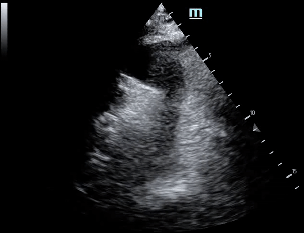
Parasternal long axis (PLAX) - the descending aorta is seen just deep to the LA. The aortic root can also be measured in this view (>4cm is abnormal)
Parasternal short axis (PSAX) - a slightly modified PSAX view by fanning the transducer may reveal the descending thoracic aorta in long axis.
Case resolution
The patient was moved to the Resus room and started on a labetalol infusion pending CT. CT aortogram confirmed a type B dissection distal to the left subclavian vessels which was medically managed (analgesia, hypertensive control, lower limb vascular monitoring) in the high dependency unit.
Take-Home Message
CT aortogram is still the gold standard for diagnosing thoracic aortic syndrome (dissection, penetrating aortic ulcer, intramural haematoma).
Bedside-focused Transthoracic echo has a high specificity for Type A and B dissection, moderate to high sensitivity for Type A but low for Type B, and is unclear for penetrating aortic ulcer and intramural haematoma. D-dimer as a blood biomarker has a high specificity, and clinical decision tool such as ADD-RS has a high sensitivity in low risk case. An approach that combines all three tests might help stratify which patients requires CT (currently there is only a low yield of positive scans)